
Medical component grinding demands more than tight dimensional tolerance and low surface roughness. For implant-grade alloys including Ti-6Al-4V, 316LVM stainless steel and CoCrMo, thermal and mechanical loading during abrasive grinding generates a thin, microstructurally altered subsurface feature widely known as the white layer. Often invisible to visual inspection, this brittle nanocrystalline zone can degrade fatigue life, corrosion resistance and biocompatibility of surgical implants and instruments. For manufacturers working under ISO 13485, EU MDR and FDA 21 CFR Part 820, identifying, quantifying and eliminating white layer formation is not merely surface-finish refinement but core risk-control activity aligned with ISO 14971 medical device risk management.
What Is the White Layer in Medical Grinding?
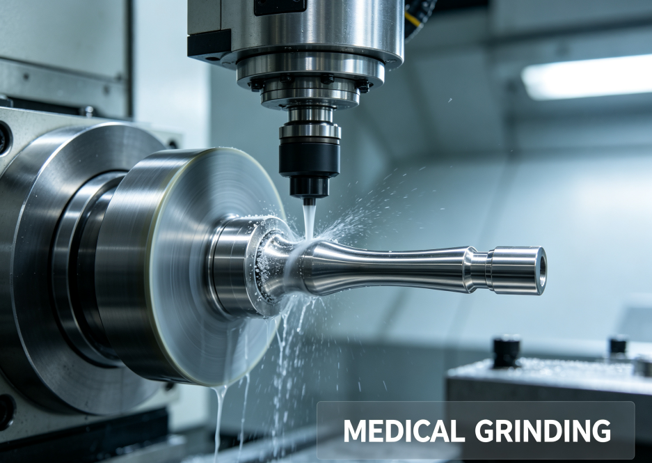
The white layer is a near-surface metallurgically altered zone that appears bright and featureless under optical microscopy after standard chemical etching, the trait that gives the feature its common name. Conventional etchants readily reveal grain boundaries in bulk substrate metal but fail to resolve microstructure within this thin band, creating stark visual contrast against underlying base material. Research published in PMC identifies two dominant formation pathways during grinding: rapid phase transformation from localized grinding heat, and severe plastic deformation with dynamic recrystallization of surface grains.
In thermally driven formation, high heat flux at the grinding wheel-workpiece interface raises surface temperature above the material’s austenitization threshold. The ultra-fast quenching effect from coolant transforms newly formed austenite into hard, brittle secondary martensite mixed with residual austenite and fine carbides, creating a hardened subsurface layer with mechanical properties markedly different from the bulk alloy. For titanium implant alloys, the second mechanism is more prevalent: extreme shear deformation fragments base grains down to nanoscale dimensions without full phase transformation, forming the same optically white, refined microstructure. Many grinding processes create white layers from a combination of both thermal and mechanical effects.
White layer thickness varies heavily with process parameters. PMC studies on hardened alloy steel show higher wheel speed, excessive feed rates and insufficient coolant delivery increase peak contact temperature and push white layer depth above 50 μm under aggressive grinding conditions. In medical-grade titanium alloys, even seemingly conservative grinding parameters can generate layers ranging from 1 μm to 20 μm. Unlike grinding burn discoloration, white layer may form without visible surface oxidation marks. This creates a hidden failure risk: a part passing dimensional check and surface roughness inspection can still carry subsurface damage undetectable by standard shop metrology.
Patient and Device Risks of Uncontrolled White Layers in Medical Implants
The mechanical and biological risks tied to white layers carry particular weight for load-bearing implant components such as bone screws, spinal fixation plates, hip and knee articulating surfaces. The layer itself exhibits significantly higher hardness and much lower ductility than the parent substrate. Internal vacancy clusters and microdefects within the white layer act as crack initiation sites under cyclic physiological loading. Published metallurgical data confirms grinding-induced white layers can reduce low-cycle fatigue life by nearly 30 times in titanium alloy components. For orthopedic implants designed to withstand decades of repeated body load, this degradation directly raises risk of in-vivo fracture and early implant revision surgery. PMC retrieval studies of hip implant taper interfaces demonstrate that surface microcracks originating from subsurface damage propagate under cyclic micromotion, ultimately leading to fatigue fracture of Ti-6Al-4V modular implant components.
Corrosion performance represents a second major hazard. The nanoscale microstructure and residual tensile stress trapped within the white layer disrupt the native passive oxide film that gives titanium and 316L stainless steel their biocompatibility. In the saline, protein-rich environment of human tissue, chloride ions easily penetrate damaged passive films, increasing susceptibility to pitting corrosion and crevice corrosion. Corrosion breakdown releases metallic ions into surrounding tissue. For cobalt-chromium-molybdenum and titanium alloys, elevated ion release triggers local inflammatory response, osteolysis, hypersensitivity reactions and premature loosening of the implant-bone interface. While bulk material meets ASTM biocompatibility standards, the modified white layer surface cannot retain the stable electrochemical behaviour required for long-term implantation.
Residual stress adds further complexity. Grinding-induced white layers almost always carry non-uniform residual stress fields. Tensile residual stress at or just below the surface propagates microcracks during sterilization cycling and physiological loading. Compressive stress can form in adjacent subsurface zones, creating unstable stress gradients that shift over time after implantation. These gradients may distort tight geometric features post-processing, causing loss of taper fit or thread engagement in modular implant systems. ISO 14971 risk assessment protocols classify this as a high-severity failure mode for permanently implanted devices, requiring formal process validation to prevent occurrence.
Beyond mechanical and corrosion risks, white layer can interfere with osseointegration. Dental and orthopedic implants rely on controlled, reproducible surface topography for bone ingrowth. The brittle white layer may spall into micro-flakes once implanted, leaving loose metallic particulate debris in the bone-implant gap. A PMC systematic review on titanium implant particle release confirms these particles activate macrophage inflammatory pathways, inhibiting bone formation and accelerating peri-implant bone resorption and implant failure. Even thin white layers alter surface chemistry enough to change protein adsorption at the material-tissue boundary, shifting biological response away from stable osseointegration.

Detecting White Layer in Medical Grinding Quality Control
Because white layer often lacks visual signs, medical manufacturing quality systems require targeted metallurgical verification, not only conventional dimensional inspection. The gold standard method is cross-sectional metallographic sample preparation: sectioning, mounting, polishing and chemical etching of sacrificial witness coupons machined alongside production parts. Under optical microscopy at 500× to 1000× magnification, inspectors measure white layer thickness and check for associated microcracks. This destructive method is reliable but cannot run on every finished implant part, so ISO 13485 mandates process validation using coupon testing to prove parameters consistently limit or eliminate white layer formation.
Complementary techniques support quantitative characterization. Microhardness traverses across the subsurface detect the sharp hardness spike characteristic of white layer. Transmission electron microscopy (TEM) enables nanoscale observation of grain refinement and phase composition, ideal for root-cause analysis when non-conformance is identified. Focused ion beam (FIB) sectioning can isolate small surface regions for high-resolution imaging without full component destruction, though cost limits routine use on production batches.
Non-destructive screening methods remain an active research area but stay secondary for regulated implant manufacturing. Acoustic emission and eddy current techniques can flag thermal damage, yet they lack sufficient resolution to quantify sub-micron white layer thickness for FDA or notified-body audit requirements. For medical component production, process validation remains the primary control strategy: once validated grinding parameters are locked in, batch release relies on process record review plus periodic destructive coupon verification.
White Layer Removal Strategies for Medical-Grade Components
When white layer is detected on machined medical parts, removal must preserve critical dimensional tolerances, surface integrity and material biocompatibility. The chosen post-processing method must not introduce new surface contamination, residual stress or geometric deviation, and the full process must be validated under ISO 13485 section 7.5.2 for special processes whose results cannot be fully verified by final inspection.
Fine abrasive polishing and controlled mass finishing are widely used for low-thickness white layer removal. Precision ceramic tumbling media with neutral pH compound gently removes the altered subsurface without aggressive material stock removal. For Ti-6Al-4V and CoCrMo implants, validated ceramic media removes 1–5 μm of surface material, stripping the white layer while preserving the engineered surface texture required for osseointegration. Careful media selection is critical: iron-containing media will contaminate implant surfaces and compromise corrosion resistance, so medical-grade finishing lines use alumina or silicon carbide media certified free of cross-contaminating trace metals.
Electropolishing provides another robust removal route, particularly for 316LVM stainless steel cardiovascular and surgical instrument components. Electropolishing selectively dissolves the high-stress, altered surface layer, removing white layer while simultaneously generating a uniform, passive chromium-rich oxide film. The process lowers surface roughness and relieves near-surface tensile residual stress, delivering dual benefits for corrosion resistance and fatigue performance. Electropolishing must be tightly controlled for material removal rate to avoid exceeding drawing tolerances, and full bath validation and bath lot traceability are required for implant applications.
Micro-grinding with optimized parameters can remove thicker white layers, but this carries risk of reintroducing new thermal damage. If re-grinding is required, shops use ultra-fine-grain, well-dressed grinding wheels, reduced depth of cut, lower wheel speed and high-volume flood coolant formulated for medical machining. The objective is minimal heat generation during stock removal, preventing a secondary white layer from forming. This rework path demands separate process validation and full metallographic confirmation after rework.
Chemical etching is rarely used as a standalone white layer removal step for implantable parts. While selective etchants can attack the altered surface layer, over-etching creates unpredictable surface topography, pitting and dimensional loss. It also introduces risk of residual etchant entrapment in micro-features, which can lead to cytotoxic residues after sterilization. Where chemical treatment is used, it is paired with passivation and multi-stage ultra-pure water rinsing, documented and validated as part of the full finishing workflow.
Preventing White Layer: Process Controls for Medical Grinding
The most cost-effective and regulatory-compliant strategy is preventing white layer formation before it occurs, rather than removing it post-grinding. PMC research confirms white layer formation is highly sensitive to grinding temperature, making thermal management the core control lever. Process controls start with wheel selection and maintenance. Vitrified-bond, fine-grain, sharp diamond or cubic boron nitride wheels are preferred for medical alloy grinding. Dull or loaded grinding wheels increase friction and localized heat, drastically raising white layer risk. Regular wheel dressing maintains cutting edge sharpness and prevents smearing of workpiece material across the surface.
Coolant selection and delivery are equally critical. Medical grinding uses chlorine-free, low-residue synthetic coolant, delivered at sufficient flow rate and pressure to flood the grinding contact zone continuously. Coolant serves not only to remove heat but also evacuate fine chips and prevent wheel loading. Coolant filtration must remove submicron abrasive particles to avoid embedded contamination on implant surfaces. For titanium alloys, inadequate cooling creates compound risk, as high temperature can simultaneously generate white layer and alpha-case embrittlement, a separate surface defect unique to titanium implant materials.
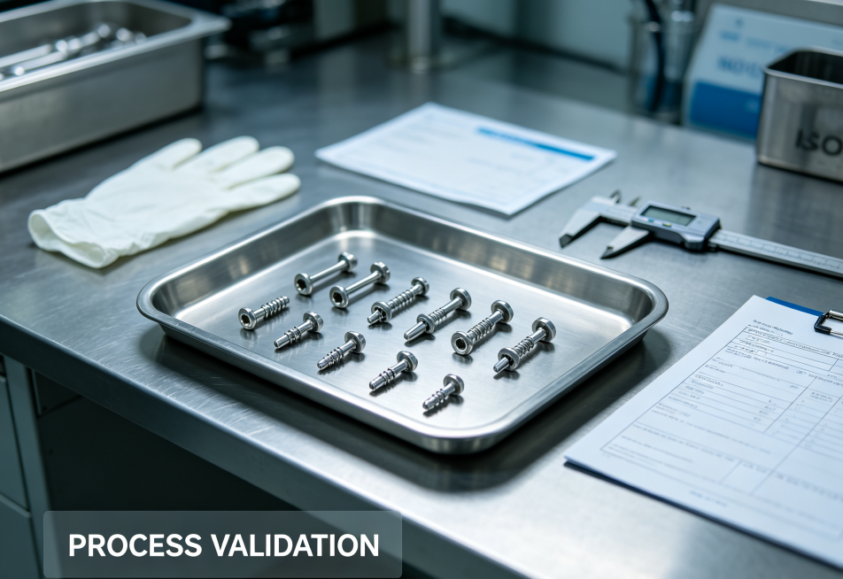
Grinding parameter validation is required under ISO 14971 risk management. Manufacturers perform design of experiments (DOE) to map how wheel speed, feed rate, depth of cut and contact time affect subsurface metallurgy. The validated process window keeps peak grinding temperature below the material’s phase transformation threshold and minimizes plastic shear strain. Once a safe operating window is defined, CNC programs lock parameters, and process records document every batch for full traceability. In-process temperature monitoring, power draw tracking and periodic coupon metallography confirm the process remains within validated limits through production runs.
Fixturing and part support also reduce white layer risk, especially for thin, delicate medical components such as cranial plates or thin-walled catheter housings. Poor fixturing creates chatter and localized pressure spikes, concentrating heat in small surface zones. Custom low-contact, non-marring fixtures distribute clamping force evenly, reducing workpiece deflection and inconsistent cutting loads.
Quality Assurance and Regulatory Considerations
Regulators treat white layer as a surface integrity failure mode for implantable medical devices. Under EU MDR 2017/745 and FDA 21 CFR Part 820, manufacturers must document risk analysis, process validation and acceptance criteria for surface microstructure. Acceptance limits for white layer thickness are defined by device risk class, material and clinical function. Many orthopedic implant specifications mandate zero detectable white layer on load-bearing implant surfaces, with validation performed using metallographic coupons.
Process validation follows the standard IQ/OQ/PQ framework. Installation qualification verifies machine, wheel, coolant and fixturing are installed correctly. Operational qualification tests parameter boundaries to define safe operating limits, including upper and lower bounds for feed, speed and coolant flow. Performance qualification runs multiple production batches with witness coupons to demonstrate consistent control of white layer thickness over sustained production. All metallographic reports, material certificates, coolant logs and SPC data form part of the device’s technical file, available for notified-body or FDA audit.
Ancastings Precision’s medical CNC and grinding services implement this full framework for implant-grade alloy components, combining validated grinding workflows, metallographic inspection and ISO 13485 quality controls to mitigate white layer risk from prototype development through serial production. Our engineering team reviews every medical component drawing to define surface integrity acceptance criteria early in quoting, so white layer risks are addressed before the first part is machined, not discovered after production.
Conclusion
White layer formation remains one of the most overlooked yet consequential failure modes in medical grinding. This thermally and mechanically altered subsurface layer can reduce fatigue performance, accelerate corrosion, release metallic ions and disrupt osseointegration, creating tangible risk for patients receiving surgical implants. Detection requires metallographic testing, as visual inspection and standard roughness measurement cannot reliably identify the defect. While multiple post-processing techniques remove white layer, validated process controls to prevent its formation deliver superior regulatory compliance, lower scrap rates and consistent component performance.
For medical device developers and OEMs, supplier selection must include verification of surface integrity controls beyond simple tolerance capability. A precision manufacturer with formal white layer risk assessment, metallurgical validation and ISO 13485 traceability is essential for implant-grade grinding. By designing grinding processes to limit heat and plastic deformation, medical component manufacturers protect patient safety while meeting the stringent requirements of global medical device regulations. If you need support with process validation, metallurgical inspection or medical-grade grinding for your next implant or surgical instrument project, contact our engineering team to review your component requirements.